Imagine catching a serious illness before it even causes symptoms, giving you the upper hand in treatment and recovery. That’s the power of diag image technology today. With advancements in diagnostic imaging, early disease detection has become more precise and accessible, potentially boosting survival rates for conditions like breast cancer to as high as 98 percent when spotted early. As we dive into this topic, we’ll explore how these tools are revolutionizing healthcare, making it easier for everyone from patients to doctors to stay ahead of health challenges.
What is Diag Image?
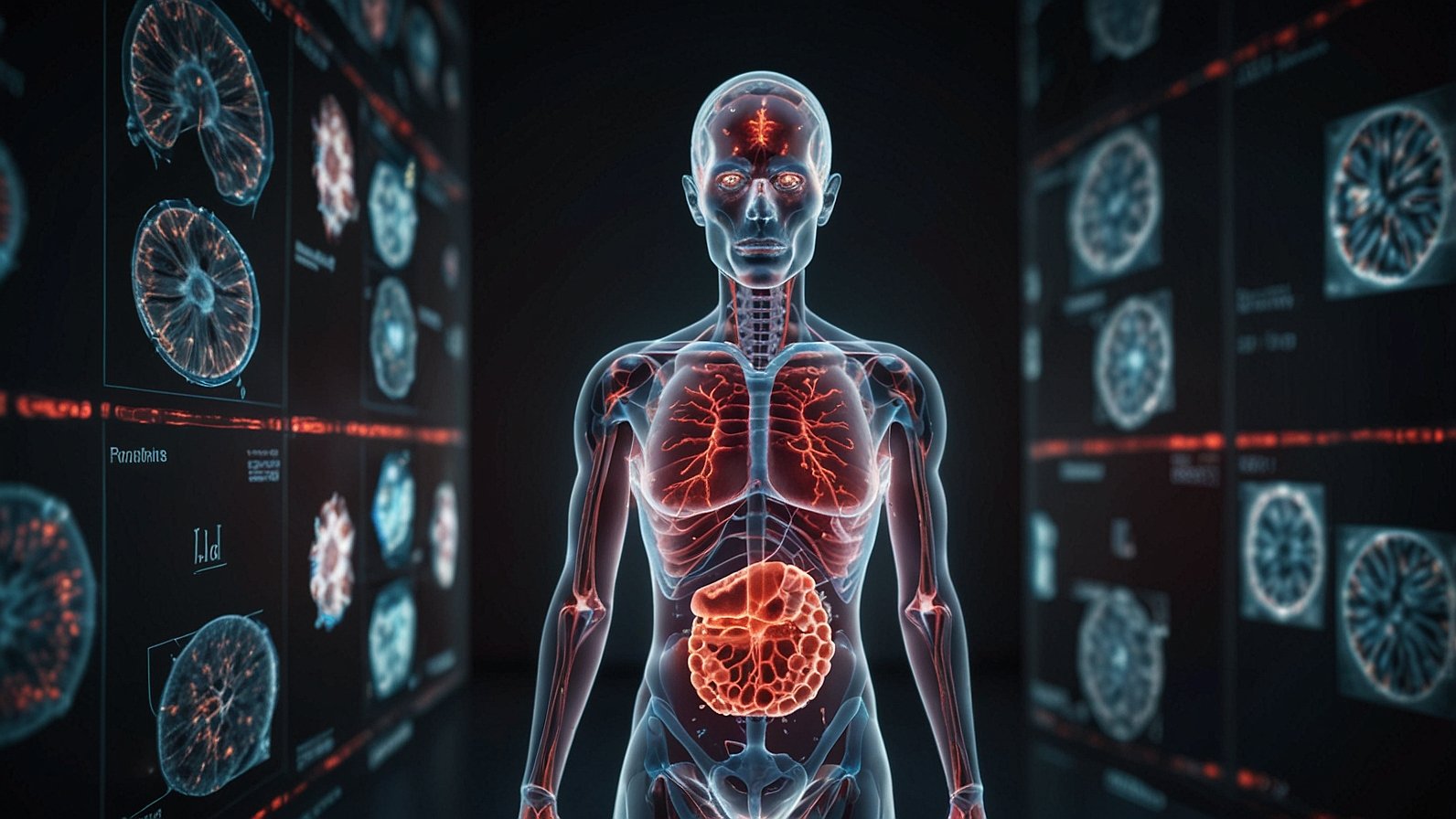
Diag image, short for diagnostic imaging, refers to a range of non-invasive procedures that let doctors peek inside the body without surgery. Think of it as a high-tech window revealing internal structures like bones, organs, and tissues. These scans use various energies, such as sound waves or magnetic fields, to create detailed pictures that help identify issues early.
At its core, diag image plays a crucial role in modern medicine by aiding in diagnosis, monitoring treatments, and even guiding procedures. For instance, it can spot tumors, fractures, or blockages that might otherwise go unnoticed until they worsen. Unlike older methods that relied on guesswork or invasive tests, today’s versions are quicker and safer, often completed in under an hour.
Healthcare practitioners rely on these tools daily, while patients appreciate the reassurance they provide. If you’ve ever wondered what a diag image is used for, it’s essentially about catching problems before they escalate, improving patient outcomes across the board.
Common Types of Diag Image Modalities
Diagnostic imaging comes in several forms, each suited to different needs. Here’s a quick rundown of the most common ones, along with what they do best.
- X-rays: The classic option for viewing bones and detecting fractures or infections. They’re fast and use low levels of ionizing radiation.
- CT Scans (Computed Tomography): These provide cross-sectional views, ideal for spotting internal injuries or cancers. They combine multiple X-ray images for a 3D-like picture.
- MRI (Magnetic Resonance Imaging): Great for soft tissue details, like brain or joint issues, using magnets and radio waves instead of radiation.
- Ultrasound: Uses sound waves to image organs or monitor pregnancies. It’s portable, real-time, and radiation-free.
- PET Scans (Positron Emission Tomography): Often used in cancer screening, they highlight metabolic activity with a small amount of radioactive tracer.
To make it easier to compare, consider this table of key features:
| Modality | Best For | Uses Radiation? | Typical Duration |
|---|---|---|---|
| X-ray | Bones, chest | Yes (low) | 5-10 minutes |
| CT Scan | Internal organs, trauma | Yes (moderate) | 10-30 minutes |
| MRI | Soft tissues, nerves | No | 30-60 minutes |
| Ultrasound | Pregnancy, abdomen | No | 15-45 minutes |
| PET | Cancer, brain function | Yes (low) | 30-90 minutes |
Each type has its strengths, and doctors choose based on the suspected issue. For example, an ultrasound might be first for abdominal pain, while a CT could follow for more detail.
The Vital Role of Diag Image in Early Disease Detection
Early detection is a game-changer in healthcare, and diagnostic imaging stands at the forefront. By revealing problems like tumors or heart issues before symptoms appear, these scans can dramatically improve survival chances. Research shows that early diagnosis via imaging boosts treatment effectiveness by around 20 percent in many cases. For breast cancer, detection in the initial stages leads to a 98 percent five-year survival rate.
Take cancer screening as an example. A routine mammogram, a type of diag image, can identify breast abnormalities years before they’re palpable. Similarly, CT scans for lung cancer in high-risk smokers have reduced mortality by spotting issues early. This isn’t just about saving lives; it’s about better patient outcomes, like shorter hospital stays and less aggressive treatments.
For health-conscious consumers, understanding this role means prioritizing screenings. Tech enthusiasts might note how portable diag image devices are making these checks more accessible, even in remote areas.
How AI is Powering the Next Wave of Diag Image Innovation
Artificial intelligence is shaking up diagnostic imaging, making it smarter and faster. AI algorithms analyze scans in seconds, spotting patterns that might escape even experienced radiologists. Recent studies highlight AI achieving 94 percent accuracy in detecting tumors, often outperforming humans in speed and consistency.
Picture AI as a tireless assistant: It flags potential issues on an MRI, prioritizes urgent cases in busy hospitals, and even enhances image quality from noisy scans. Companies like Google Health and IBM Watson are leading with tools that integrate AI into radiology workflows, reducing errors and wait times.
For instance, in AI-powered diag image analysis, software from Aidoc helps detect brain bleeds on CT scans almost instantly, allowing quicker interventions. This intersection of AI and diagnostics isn’t futuristic; it’s happening now, with the market projected to grow to nearly USD 23 trillion by 2035. Healthcare practitioners gain efficiency, while patients benefit from more accurate diagnoses.
Key Benefits of Diag Image in Healthcare
The advantages of diagnostic imaging extend far beyond basic detection. Here are some standout perks:
- Non-Invasive Nature: No cuts or recovery time, unlike surgery. It’s like getting a detailed body map without the hassle.
- Improved Accuracy: High-resolution images lead to precise diagnoses, minimizing missteps in treatment.
- Better Patient Outcomes: Early spotting means less invasive therapies and higher recovery rates.
- Cost-Effective Over Time: By avoiding unnecessary procedures, it saves healthcare dollars. For example, a timely ultrasound can prevent costly complications.
- Versatility for Various Conditions: From heart disease to sports injuries, it’s adaptable.
Real-world examples abound. At facilities like Mayo Clinic, advanced imaging informatics streamline data sharing, enhancing collaboration among doctors. Contrast agents sometimes boost visibility, but risks like ionizing radiation are managed carefully.
Preparing for Your Diag Image Scan: What to Expect
Heading into a scan can feel daunting, but preparation is straightforward. Start by following your doctor’s instructions, which vary by type. For a CT scan, you might fast for a few hours if contrast is involved, while MRIs require removing metal objects like jewelry.
Arrive early to fill out forms about allergies or implants. Wear comfortable clothes, and let staff know if you’re anxious; many centers offer sedation for claustrophobic patients during MRIs. Hydration is key for some ultrasounds, but avoid caffeine before others.
Think of it as prepping for a photo shoot: The clearer the setup, the better the results. Portable diag image devices are simplifying this, allowing scans in clinics without big machines.
Decoding Diag Image Results: A Patient’s Guide
Understanding diag image results starts with knowing they’re not always straightforward. A radiologist interprets the images, noting abnormalities like shadows or densities. Your doctor then explains in simple terms, perhaps comparing it to a roadmap highlighting detours.
Common myths? Not every spot means cancer; many are benign. AI tools are helping here, providing second opinions for accuracy. If results show soft tissue issues on an MRI, follow-up tests might confirm. Always ask questions, and request copies for your records.
Emerging Trends Shaping the Future of Diag Image
Looking ahead, diagnostic imaging is evolving rapidly. AI integration tops the list, with tools predicting disease risks from scans. Cloud-based systems allow seamless sharing, while 3D printing from images aids surgical planning.
Wearable tech, like smart ultrasound patches, could make monitoring continuous. Expect growth in PET and CT exams by 15-23 percent in coming years. These shifts promise faster, more personalized care.
In summary, diag image technology is a lifeline in early disease detection, blending tradition with innovation for better health. To get started, schedule regular screenings based on your risk factors, discuss options with your doctor, and stay informed about AI advancements. What’s your take on these tools? Share in the comments below.
You May Also Like: Geekzilla T3 Review: Why Professionals and Gamers are Swapping Their Gear
FAQs
What is a diag image used for?
It’s primarily for visualizing internal structures to diagnose conditions early, monitor treatments, and guide procedures without invasion.
What are the benefits of diag image in healthcare?
Key perks include non-invasive detection, improved accuracy, and better patient outcomes, often reducing the need for surgery.
How does diag image help with cancer screening?
Modalities like PET and mammograms spot tumors early, increasing survival rates significantly.
What preparation is needed for a diag image scan?
It depends on the type; fasting for CTs or removing metals for MRIs are common steps.
How can I understand my diag image results?
Your doctor will explain, but ask for clarifications and consider AI-assisted reviews for added precision.
What role does AI play in diag image analysis?
AI enhances speed and accuracy, detecting issues like tumors with high reliability.
Are there portable diag image devices available?
Yes, emerging tech like handheld ultrasounds is making scans more accessible outside hospitals.